Article Science Technology

Analysing cell imaging data at scale with AI

Researchers from the Earlham Institute and The Alan Turing Institute are using AI to examine the relationship between a cell’s structure and function.

18 February 2025

The human body is made up of hundreds, if not thousands, of different types of cells. Understanding how individual cells work is a key focus for the Earlham Institute’s Cellular Genomics research programme.

Recent advances mean it’s now possible to make detailed studies of just one cell, to understand how it works, and chart how it responds to changes, such as those caused by ageing, diet, or inflammation.

One of these techniques is imaging flow cytometry, where an individual cell can be separated and analysed with the help of laser beams. This gives a better idea of how the shape and size of a cell relates to its function in the body. 

As part of its single-cell and spatial analysis platform, the Earlham Institute was an early adopter of a spectral imaging cell sorter called the BD FACSDiscover S8. 

This cutting-edge flow cytometer not only separates single cells, but it also creates thousands of images of individual cells in just one second.

But it’s a technique that produces vast amounts of data, particularly the cell images, that need to be catalogued and understood.

Dr Andrew Goldson, Platform Manager, using the BD FACSDiscover imaging cell sorter in single-cell labs at the Earlham Institute

Platform Manager, Dr Andrew Goldson, using the BD FACSDiscover S8 imaging cell sorter

Dealing with data

Dr Sonia Fonseca is a postdoc in the Wojtowicz Group at the Earlham Institute, where she studies blood cells; how they behave and how they can malfunction. 

“In the past we’ve been studying thousands of cells together in a single sample,” she says. 

“Using a single-cell approach means we can tease apart the complex mixtures of cells that make up humans, animals, and plants.”

Sonia’s multidisciplinary approach is essential for highlighting the extent of cellular diversity within an organism as a natural part of health.

Some variation is natural and it can be challenging to define the point at which morphological differences may suggest the existence of a subpopulation. To explore this, a combination of technologies is needed to address the sheer scale of data generated.

“With this imaging flow cytometry, we can generate a lot more data about the cells we’re studying,” says Sonia, “but we don’t have a fast and efficient way to process the data yet. 

“This creates a bottleneck that can slow down our research. We need to work with computational scientists to fully benefit from all the data we are collecting.”

Dr Sonia Fonseca and Dr James Wilsenach looking at a screen together

Dr Sonia Fonseca and Dr James Wilsenach

Sonia is collaborating with Dr James Wilsenach, associate researcher at the Earlham Institute and a postdoc at The Alan Turing Institute where he works with Professor Sebastian Ahnert

Their interdisciplinary approach brings together cellular and molecular biologists with computer scientists in a unique way.

James is using his expertise in data science to build a tool, called FlowFI, that can handle the cell data efficiently and, hopefully, remove the bottleneck.

“I work in artificial intelligence and one of the things [it] has been good at in the last few years is analysing images,” says James. 

“AI can examine thousands of images in a fraction of the time it would take a human to do the same.

“I think there's something really interesting we can discover by getting machines to learn about the connection between cell shape, genotype, and function.”

I think there's something really interesting we can discover by getting machines to learn about the connection between cell shape, genotype, and function.

Dr James Wilsenach

Megakaryocytes visualised from a cell sorter

A group of megakaryocytes imaged by the BD FACSDiscover S8 cell sorter

Rare and unusual cells

FlowFI works by taking a whole bunch of imaging parameters and giving a ranking to them, helping the user to understand what's in front of them.

It can be an invaluable way of identifying any subpopulations that might otherwise be missed, which is important with cell types where the cell morphology is known to be more fluid.

Working together, Sonia and James are using FlowFI in conjunction with imaging flow cytometry to study cells that are rare in the human body and unusual compared to other cell types. 

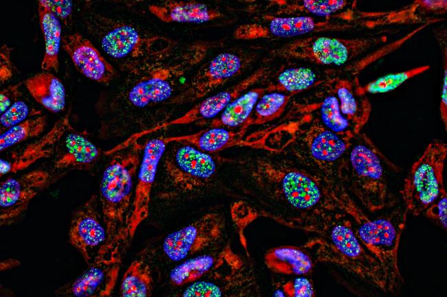
For example, Sonia is studying a type of bone marrow cell called a megakaryocyte. As the name suggests, these are far larger than most other cells found in the human body. 

Megakaryocytes are known to produce the platelets that help blood to clot, but some studies suggest that they might have other jobs in the body.

They can also increase copies of their genetic material up to 128 times, where most cells have only two copies. 

“Although megakaryocytes are normally found in the bone marrow, there’s growing evidence that they can be found in lungs and other tissues, and I’d like to know what they’re doing there,” explains Sonia. 

“But because they are bigger and more fragile than other cells, they're quite difficult to work with.”

James and Sonia viewing megakaryocytes on screen imaged by the spectral cell sorter

James and Sonia viewing megakaryocytes on screen imaged by the spectral cell sorter

With this imaging flow cytometry, we can generate a lot more data about the cells we’re studying, but we don’t have a fast and efficient way to process the data yet. This creates a bottleneck that can slow down our research. We need to work with computational scientists to fully benefit from all the data we are collecting.

Dr Sonia Fonseca

Form follows function

Sonia is using advanced flow cytometry to sift out individual megakaryocyte cells, then employing FlowFI to process large-scale imaging and genomic data together.

Ultimately, she is asking detailed questions about what a particular cell is doing - a key strand of the Cellular Genomics research programme. For example, aside from making platelets, could these cells also be doing some work for the body’s immune system?

James believes FlowFI could be applied in many other ways, including studying the weird and wonderful shapes of bacteria that can either damage or promote human health, or the beautiful crystalline structure of pollen from important crop species. 

It could eventually be used in hospitals, where flow cytometry has a role in diagnosing leukaemia.

“FlowFI is an open-source software that can work alongside existing commercial software, and it’s being developed specifically to help scientists answer questions about the structure and function of these tiny entities,” James adds.

“We want more scientists to use FlowFI because their feedback will be invaluable to refining this new tool. 

“That’s the nice thing about producing a technology like this – there are other people who are going to take up the mantle and use it for something else.”

Authored by Kerry Noble, writing for the Earlham Institute.