New research from the Earlham Institute and University of Washington provides a proof-of-principle for the recorder in accurately tracking gene expression, with a wide range of potential future applications.
The findings could one day lead to ways of engineering plant features to help improve their efficiency - particularly valuable in the context of climate change.
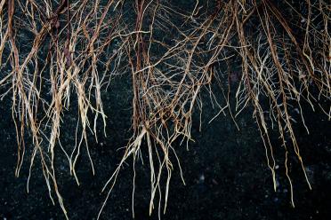
It may lie hidden beneath the ground but the intricate architecture of a plant’s root network is a critical feature. Roots help to anchor a plant, draw up water and nutrients, and provide an interface with the soil microbiome.
In a germinating plant, the primary root pushes down into the earth. As it descends, new lateral roots begin to emerge, extending outwards and splitting off into a web of smaller roots.
Many genes responsible are known but the order of processes controlling when and where lateral roots will appear is difficult to map. Understanding the step-by-step development of these roots could be used to help engineer more efficient or better-adapted structures.
Researchers at the University of Washington and the Earlham Institute wanted to develop a robust and repeatable technique for recording, at the cellular level, the order in which genes of interest are expressed.
Dr Sarah Guiziou, author and Group Leader at the Earlham Institute, said: “Most of the techniques we currently use give a snapshot of gene expression, which lacks the temporal and spatial information we need, or they require prohibitively expensive set-ups to allow live imaging.
“We wanted to construct a DNA recorder that would be able to document both the timing and location of gene expression in plants.”
Using Arabidopsis thaliana as the model for their experiments, the researchers built an integrase-based recorder - a DNA construct incorporated into the plant - engineered to cause fluorescence of different colours in plant cells depending on the expression, and order of expression, of two genes of interest.
This allowed the researchers to use imaging techniques to look for fluorescence as a hallmark of gene expression, with colour changes mediated by a DNA switch - signalling the expression of one of the genes of interest.
As the DNA switches were irreversible, both the cells and their descendents were identifiable from the same fluorescent marker, allowing the researchers to track the cell lineages.
By imaging the root once, they could see when and where their target genes were being expressed, signalling the development of a new lateral root.
“Our integrase-based recorder was able to clearly show the order of gene expression at a single-cell level, with images showing when and where lateral root development was occuring,” said Dr Guiziou.
Dr Jennifer Nemhauser, author and Professor of Developmental Biology at the University of Washington, said: “We wanted to create a way of recording the timeline of gene expression in these plants, which allows us to visualise where those genetic switches occur that lead to root development.”
The researchers also chose to investigate the development of stomata - pores found in the surface of leaves that open and close to allow oxygen to flow out and carbon dioxide to be captured from the air. The stomata are typically formed by asymmetric cell division, creating a pair of ‘guard cells’.
“We noticed some unexpected variations in the output of our DNA recorder, with part of the stomata fluorescing and others not,” explained Cassandra Maranas, a graduate student in the Nemhauser lab group at the University of Washington.
“While we were a bit surprised at first, we realised this disparity correlated with two different mechanisms for stomata development where the time between the expression of our two genes of interest differed. Our recorder was capturing the variation in timing of gene expression.”
The researchers now believe their recorder could be adapted and applied to answer a range of different questions - both in plants and animals.
Funding for the study came from the National Institutes of Health, the National Science Foundation, the Howard Hughes Medical Institute Faculty Scholars Program, and also the UKRI Biotechnology and Biological Sciences Research Council.
‘A history-dependent integrase recorder of plant gene expression with single cell resolution’ is published in the journal Nature Communications.
